A current paper revealed in Nature Growing old dives into the gene expression variations between younger, middle-aged, and older human ovaries and tests possible interventions to slow down their aging processes [1].
An underexplored space of human ageing
Feminine reproductive ageing stays a comparatively unexplored space of examine. With folks residing longer and females suspending childbearing, there’s a want for analysis into slowing it down.
The ovaries are indispensable organs in feminine copy, important in hormone manufacturing and fertility. [2] In comparison with different organs, ovarian functioning ceases comparatively early in life, at round 50 years. Nonetheless, the decline in its operate begins round 20 years earlier, when ladies attain across the age of 30 [3].
Age-dependent variations in ovaries
On this new examine, the researcher centered on exploring human ovaries and the adjustments to their gene expression (the transcriptome) with age.
One of many greatest challenges to understanding adjustments in gene expression in human ovarian ageing is acquiring tissues for research. Nonetheless, these researchers had been capable of acquire the ovaries of 15 volunteers who donated them following surgical procedure. These had been divided into three teams based mostly on age: younger (18-28 years), middle-aged (36-39 years), and older (47-49 years).
Human ovaries are organs which are constructed from several types of cells [4]. Every cell sort has completely different traits and expresses completely different genes. To grasp the ageing processes of those completely different cell varieties, the authors used strategies that measure gene expression on the single-cell stage and evaluate the gene expression between completely different age teams and cell varieties [5].
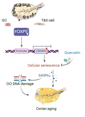
Whereas there have been many variations between every cell sort, the gene expression evaluation typically revealed “important variations between perimenopausal ovaries and people with reproductive operate in younger or middle-aged stage.” A extra detailed evaluation factors to mobile senescence as an essential participant in ovarian ageing. The authors additionally spotlight options such because the senescence-associated secretory phenotype (SASP), elevated mobile ageing hallmarks in aged ovaries, resembling SA-β-gal exercise and lipofuscin accumulation, and oxidative protein harm.
The SASP molecules secreted by senescent cells are identified to result in irritation [6]. These beforehand noticed connections had been additionally noticed on this analysis, because the researchers noticed elevated ranges of inflammation-related molecules. A rise in irritation and SASP molecules can result in the spreading of the senescent phenotype and oocyte harm, which was supported by the remark that DNA harm methods had been additionally activated in aged oocytes.
FOXP1, the essential regulator
After analyzing the widespread molecular processes and gene expression in several types of ovarian cells, the researchers appeared for the transcription issue regulating these processes. They recognized “FOXP1 as a vital TF regulating mobile senescence within the ovary” and noticed that ageing decreased FOXP1 ranges.
In addition they examined FOXP1 in mice. The depletion of FOXP1 in granulosa cells led to expedited ovarian ageing, senescence-associated adjustments in gene expression patterns, elevated ranges of senescent markers, and extra granulosa cells dying by apoptosis. Nonetheless, they famous this experiment’s limitations, as flattening FOXP1 in mice can’t absolutely replicate human ovarian ageing.
Quercetin’s anti-aging impact
The authors went a step additional and examined potential anti-aging medicine and their results on the ovarian reserve in middle-aged mice and human ovarian granulosa tumor cell strains. They examined fisetin, quercetin, and dasatinib, that are already identified to have anti-aging properties [7]. First, testing in human cell strains confirmed that quercetin and fisetin “delayed FOXP1 gene silencing-induced mobile senescence,” improved cell proliferation, and decreased ranges of a DNA harm marker. Quercetin additionally activated FOXP1 expression, suggesting that it will possibly inhibit senescence.
These promising outcomes prompted researchers to check quercetin in reproductively aged (36-week-old and 48-week-old) mice. These ages correspond roughly to 38-40 and 48-50 in people. Mice obtained one month of quercetin therapy with none negative effects. Comply with-up testing confirmed enhancements within the ovarian reserves of 36-week-old, however not 48-week-old, mice.
Moreover, quercetin-treated 36-week-old mice had been capable of carry extra profitable pregnancies in comparison with the management group. The shortage of enchancment noticed within the 48-week-old mice might be because of the restricted variety of follicles, the sacs that include immature eggs, in these outdated mice.
Total, this examine contributed to a greater understanding of feminine ageing processes. The great evaluation carried out by the authors allowed them to create “single-cell and spatial transcriptomic maps of human ovaries throughout completely different age teams.” These maps allowed for inspecting gene expression adjustments and variations in eight forms of human ovarian cells.
Our examine deepens the understanding of human ovarian ageing, offering a helpful useful resource for investigating potential therapeutic interventions. Transferring ahead, we goal to discover FOXP1 as a possible goal for each the analysis and therapy of human ovarian ageing.

Literature
[1] Wu, M., Tang, W., Chen, Y., Xue, L., Dai, J., Li, Y., Zhu, X., Wu, C., Xiong, J., Zhang, J., Wu, T., Zhou, S., Chen, D., Solar, C., Yu, J., Li, H., Guo, Y., Huang, Y., Zhu, Q., Wei, S., … Wang, S. (2024). Spatiotemporal transcriptomic changes of human ovarian aging and the regulatory role of FOXP1. Nature ageing, 10.1038/s43587-024-00607-1. Advance on-line publication.
[2] Baerwald, A. R., Adams, G. P., & Pierson, R. A. (2012). Ovarian antral folliculogenesis during the human menstrual cycle: a review. Human copy replace, 18(1), 73–91.
[3] Broekmans, F. J., Knauff, E. A., te Velde, E. R., Macklon, N. S., & Fauser, B. C. (2007). Female reproductive ageing: current knowledge and future trends. Tendencies in endocrinology and metabolism: TEM, 18(2), 58–65.
[4] Hsueh, A. J., Kawamura, Ok., Cheng, Y., & Fauser, B. C. (2015). Intraovarian control of early folliculogenesis. Endocrine evaluations, 36(1), 1–24.
[5] Tabula Muris Consortium (2020). A single-cell transcriptomic atlas characterizes ageing tissues in the mouse. Nature, 583(7817), 590–595.
[6] Muñoz-Espín, D., & Serrano, M. (2014). Cellular senescence: from physiology to pathology. Nature evaluations. Molecular cell biology, 15(7), 482–496.
[7] Di Micco, R., Krizhanovsky, V., Baker, D., & d’Adda di Fagagna, F. (2021). Cellular senescence in ageing: from mechanisms to therapeutic opportunities. Nature evaluations. Molecular cell biology, 22(2), 75–95.


